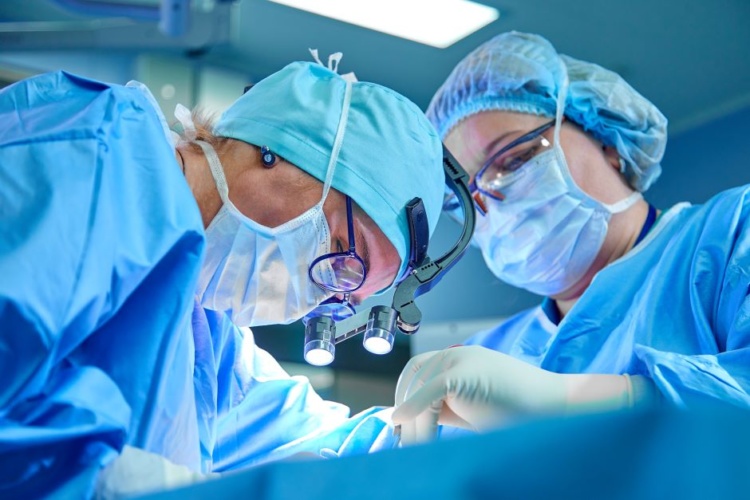
The robot-assisted surgery technique – pioneered by the National Robotarium, industry partners and Edinburgh-based clinicians - has been awarded £1.25m by the Engineering and Physical Sciences Research Council. It will be used during robotic surgery to help decide how much of the patient’s tissue is affected by cancer and should be removed. The new method will provide surgeons with real-time feedback, allowing for greater precision when differentiating normal from abnormal tissue.
The outer rim of tissue which the surgeon chooses to remove is called the ‘surgical margin’, which is identified using the surgeon's experience, preoperative imaging visual observations and, in open surgery, tactile 'feel'. Another method involves sending specimens during the operation to the pathology lab for ‘frozen section’ which takes 15-20 minutes. When undertaking ‘keyhole surgery’ using laparoscopic, endoscopic or robotic operations, surgeons can’t use 'feel' to determine tissue characteristics.
How the Versius robot could bring keyhole surgery to the masses
Carriage cleaning robot to cleanse hard-to-reach places
The new collaboration will allow mechanical measurements to be taken inside and around the surgical target which will be interpreted using a set of so-called 'mechanical intelligence' algorithms. According to the National Robotarium, the data will provide clinicians with a clear indication of a tissue’s disease status and determine how much tissue to remove during the operation.
The research team will work alongside industry partners IntelliPalp Dx and CMR Surgical along with clinicians working in the Western General Hospital in Edinburgh.
In a statement, research leader Dr Yuhang Chen, from the National Robotarium, said: “This new technique will offer surgeons a quantitative, real-time, reliable and evidence-based method for determining the optimal surgical margin to make when removing a tumour.
“Surgeons operating along a ‘keyhole’ or using techniques for minimally invasive surgery need to identify different structures or diseased areas, even when these look very similar. Our work is aimed at identifying the optimum margin in cancer surgery, to allow the removal of a tumour together with enough tissue to ensure the cancer is completely removed, but without excess being lost.
“We’re bringing together expertise from laser manufacturing, fibre-optic sensors, micromechanical probing and computational modelling to create a mechanical 'imaging' probe capable of detecting cancerous tissue that can be used with a standard minimally-invasive surgery instrument. Coupled to this, we’ll be building a 'mechanically-intelligent' data modelling framework and will integrate it into the probe operation for tumour identification and surgical margin assessment. This will effectively eliminate the margin of error for surgeons, giving them confidence that they have removed the correct amount of tissue during the operation itself and reduce the need for further invasive surgery for patients.”


McMurtry Spéirling defies gravity using fan downforce
Ground effect fans were banned from competitive motorsport from the end of the 1978 season following the introduction of Gordon Murray's Brabham...