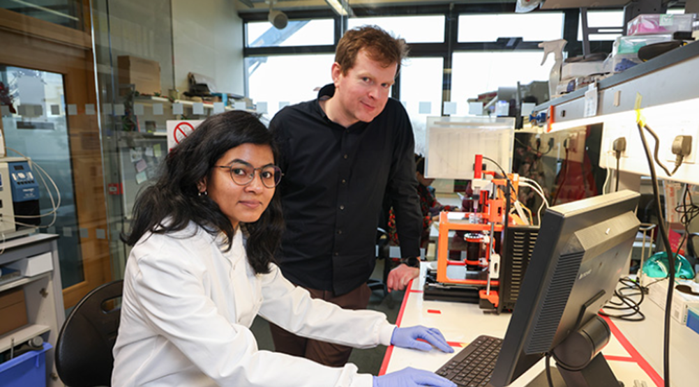
Focusing on heart tissue, the study showed that cell-generated forces could guide the shape-morphing of bioprinted tissues. The Galway team also demonstrated that tweaking factors such as the initial print geometry and bioink stiffness impacted the magnitude of the shape changes, and that the morphing enhanced the functionality of the printed tissue. The work is published in Advanced Functional Materials.
Related content
“Our work introduces a novel platform, using embedded bioprinting to bioprint tissues that undergo programmable and predictable 4D shape-morphing driven by cell-generated forces,” said lead author Ankita Pramanick, CÚRAM PhD Candidate at Galway University.
“Using this new process, we found that shape-morphing improved the structural and functional maturity of bioprinted heart tissues.”
To date, bioprinting methods have generally sought to directly replicate the final anatomical shape of organs such as the heart. According to the researchers, this doesn’t leave room for the crucial role of dynamic shape changes that take place during natural embryonic development. In the heart’s case, it begins as a simple tube that then undergoes a series of bends and twists to form its mature four-chambered structure. Facilitating this cell morphing could be a vital step towards printing functional human organs.

“Our research shows that by allowing bioprinted heart tissues to undergo shape-morphing, they start to beat stronger and faster,” said principal investigator Andrew Daly, Associate Professor in Biomedical Engineering at Galway University.
“The limited maturity of bioprinted tissues has been a major challenge in the field, so this was an exciting result for us. This allows us to create more advanced bioprinted heart tissue, with the ability to mature in a laboratory setting, better replicating adult human heart structure.”
But according to Daly, in vitro applications are still some way off. For the technique to replicate organ function, additional components such as blood vessels would need to be incorporated into the bioprinted tissue.
“We are still a long way away from bioprinting functional tissue that could be implanted in humans, and future work will need to explore how we can scale our bioprinting approach to human-scale hearts,” said the Professor.
“We will need to integrate blood vessels to keep such large constructs alive in the lab, but ultimately, this breakthrough brings us closer to generating functional bioprinted organs, which would have broad applications in cardiovascular medicine.”


McMurtry Spéirling defies gravity using fan downforce
Ground effect fans were banned from competitive motorsport from the end of the 1978 season following the introduction of Gordon Murray's Brabham...