An adaption of fibre-optic sensors by researchers from the Institute of Photonics technology at Jinan University in Guangzhou has led to the development of a fibre laser photoacoustic imaging technique that exploits the effects of sound on laser pulses. The technique may have applications in wearable devices, instrumentation and medical diagnostics.
Presenting their research at an optics and laser science conference in Washington DC earlier this week, the team, led by Long Jin, explained how their technique depends on the thermo-elastic effect – temperature changes that occur as a result of elastic strain in optical fibres. According to Jin, this effect is commonly used in military fibre laser applications to detect acoustic waves in the kilohertz frequency range. "But it turns out that they don't work so well for ultrasound waves at the megahertz frequencies used for medical purposes because ultrasound waves typically propagate as spherical waves and have a very limited interaction length with optical fibers," he said. This limits their use in medical imaging, where ultrasound is frequently used. Medical ultrasound machines use piezoelectric detectors, whose sensitivity is rather low.
The Jinan team developed its sensor specifically for medical ultrasound imaging. Essentially, the sensor is a very compact laser, built within the core of a single-mode optical fibre with diameter of around 8 µm. To build the laser, the team "wrote" two highly-reflective grating mirrors into the fibre core with ultraviolet light. The glass fibre was then doped with ytterbium and erbium to convert it into a laser medium, which was pumped to initiate laser activity using a 980 nm semiconductor laser. The fibre, typically only 8 mm long, then emitted light at the 532nm wavelength.
When these fibre lasers are impacted by ultrasound waves parallel to the length, the fibre deforms, which modulates the emitted frequency of light. "By detecting the frequency shift, we can reconstruct the acoustic waveform," explained research team member Yizhi Liang.
To reconstruct the waveform, the team uses a method called self-heterodyning, which involves measuring the effect of the mixing of two frequencies. This has the advantage over a more common method, using interferometry because the signal output is more stable.
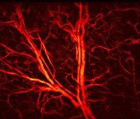
The team tested the laser by studying the blood vessels in a mouse's ear, using a focused 532-nanometer nanosecond pulse laser. “This method can also be used to structurally image other tissues and functionally image oxygen distribution by using other excitation wavelengths -- which takes advantage of the characteristic absorption spectra of different target tissues," Jin said.
These sensors have the potential to make much narrower endoscopes, Jin suggested. “current commercial endoscopic products are typically millimetres in dimension, which can cause pain, and they don't work well within hollow organs with limited space."


April 1886: the Brunkebergs tunnel
First ever example of a ground source heat pump?