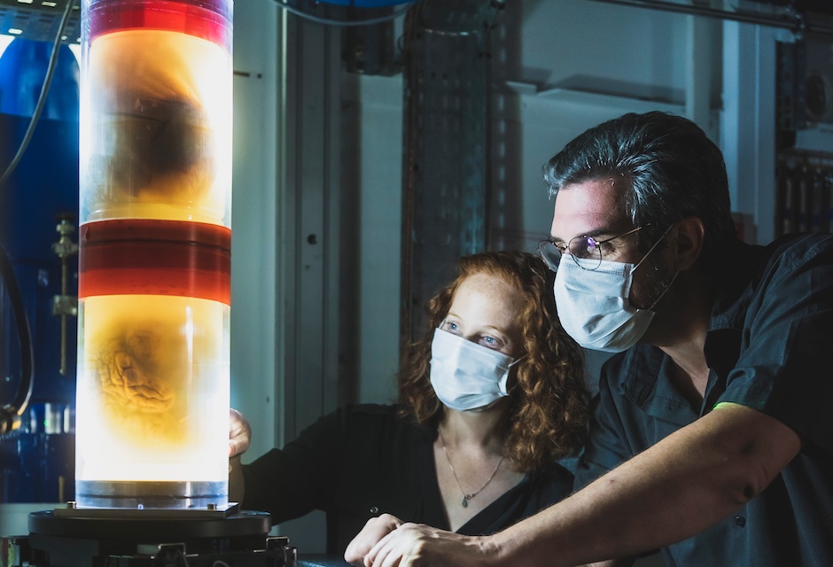
The damage to the lungs’ smallest blood vessels was captured using high-energy X-rays emitted by a special particle accelerator, which scanned a range of donated human organs including lungs from a Covid-19 donor.
HiP-CT enables 3D mapping across a range of scales, allowing clinicians to image the whole organ and then zoom down to cellular level.
The technique uses X-rays supplied by the European Synchrotron particle accelerator in Grenoble, France, which following its recent Extremely Brilliant Source upgrade (ESRF-EBS) now provides the brightest source of X-rays in the world — 100 billion times brighter than a hospital X-ray.
Due to this, researchers can view blood vessels five microns in diameter (a tenth of a diameter of a hair) in an intact human lung. A clinical CT scan only resolves blood vessels that are around 100 times larger, about 1mm in diameter.
Dr Claire Walsh, UCL Mechanical Engineering, described the technique as revolutionary for medical imaging, commenting: “As we start to link our HiP-CT images to clinical images through AI techniques, we will — for the first time — be able to highly accurately validate ambiguous findings in clinical images.
MORE MEDICAL AND HEALTHCARE NEWS HERE
“For understanding human anatomy this is also a very exciting technique, being able to see how tiny organ structures in 3D in their correct spatial context is key to understanding how our bodies are structured and how they therefore function.”
HiP-CT showed the research team, which includes clinicians in Germany and France, how severe Covid-19 infection ‘shunts’ blood between the two separate systems — the capillaries which oxygenate the blood and those which feed the lung tissue itself. Such cross-linking stops the patient’s blood from being properly oxygenated, which was previously hypothesised but not proven.
Dr Paul Tafforeau, lead scientist at ESRF said that the idea for the HiP-CT technique came after the beginning of the pandemic, by combining techniques used at the ESRF to image large fossils and using the increased sensitivity of the ESRF-EBS.
“This allows us to see in 3D the incredibly small vessels within a human organ, enabling us to distinguish in 3D a blood vessel from the surrounding tissue, and even to observe some specific cells,” he said. “This is a real breakthrough, as human organs have low contrast and so are very difficult to image in detail with the current available techniques.”
With support from the Chan Zuckerberg Initiative, the UCL-led team is using HiP-CT to produce a Human Organ Atlas. This will display six donated control organs: brain, lung, heart, two kidneys and a spleen, and the lungs of a patient who died of Covid-19.
There will also be a control lung biopsy and a Covid-19 lung biopsy. The Atlas will be available online for surgeons, clinicians and the interested public.
Project lead Professor Peter Lee, UCL Mechanical Engineering, said that the Atlas spans a ‘previously poorly explored scale in our understanding of human anatomy’ — he added that clinical CT and MRI scans can resolve to just below a millimetre, whilst histology (studying cells/biopsy slices under a microscope), electron microscopy (using an electron beam to generate images) and other similar techniques resolve structures with sub-micron accuracy but only on small biopsies of tissue from an organ.
Researchers believe the scale-bridging imaging technique could provide additional insight into many diseases such as cancer or Alzheimer’s Disease.
The authors hope the Human Organ Atlas will eventually contain a library of diseases that affect organs on a range of scales, and plan to use machine learning and AI to calibrate clinical CT and MRI scans to enhance understanding of clinical imaging and enable faster, more accurate diagnosis.
Collaboration to address viable solutions for VAWG database
<blockquote>address the lack of standardisation, coordination, and collaboration of gender disaggregated data intelligence across various regions,...